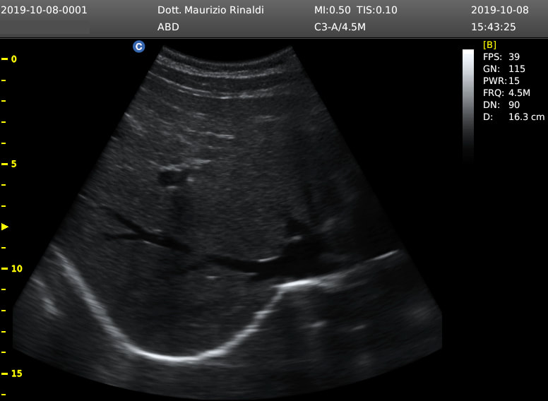
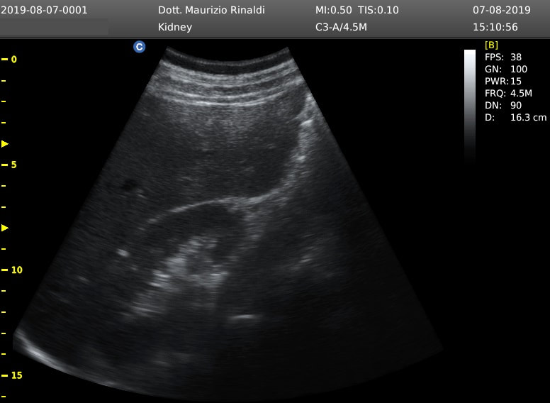
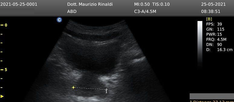
- When to perform scrotal ultrasound?
- No preparation is required before the exam.
- testicular swelling
- testicular pain
- bleeding
- infertility
- No preparation is required before the exam.


 Feed RSS