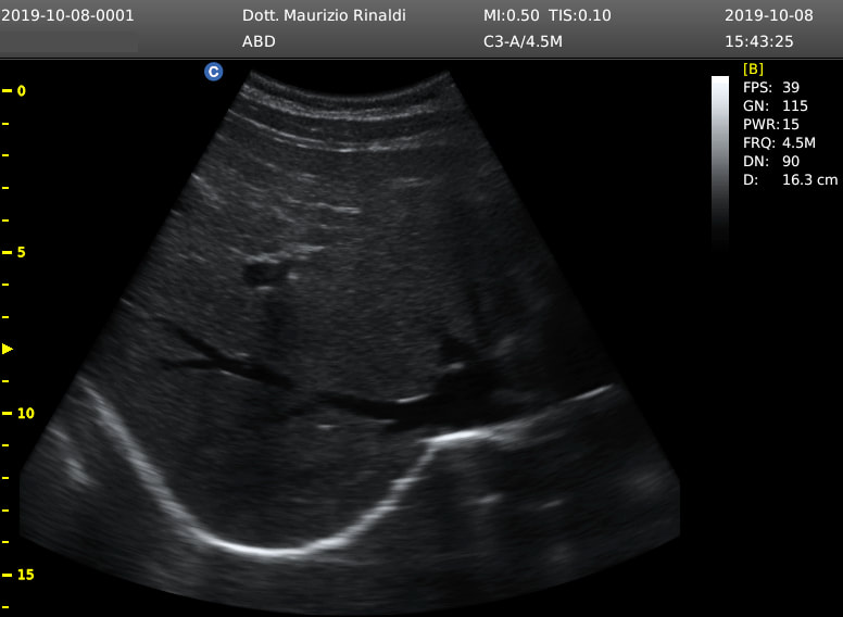
Available Ultrasound
The ultrasound is also carried out at home, by reservation, with immediate clinical report.
During the ultrasound examination, the probe of the instrument is resting on the skin surface of the patient.
An aqueous gel is smeared on the area to be studied to facilitate the transmission of ultrasound, which will allow to view the organs on a monitor connected to the probe.
Remember to bring with you any ultrasound examinations already carried out previously.
Clinical indications
> Abdominal ultrasound:
Abdominal ultrasound is a fast, painless examination without contraindications.
- What is its diagnostic utility?
Abdominal ultrasound is a diagnostic medical examination useful for studying the position, shape and any alterations of the organs contained in the abdominal cavity:
- What diagnoses does it allow?
- To reduce the air in the intestine and make the gallbladder stretch well before the examination it is necessary to fast for at least 6 hours. However, you can drink water and continue taking any medications
- What diet should you follow in the days before the examination?
2 days before the ultrasound it is advisable to follow a light diet and reduce the intake of:
Remember to drink half a liter of water after urinating two hours before the examination.
Abdominal ultrasound is a fast, painless examination without contraindications.
- What is its diagnostic utility?
Abdominal ultrasound is a diagnostic medical examination useful for studying the position, shape and any alterations of the organs contained in the abdominal cavity:
- liver
- gall bladder
- pancreas
- spleen
- kidneys
- bladder
- internal genitalia:
- What diagnoses does it allow?
- liver disease
- diseases of the gallbladder
- pancreatitis
- cysts and abscesses
- kidney disease, such as urinary tract obstructions or nephritis
- benign or malignant tumors
- presence of fluid in the abdominal cavity
- alterations of the intestine walls
- To reduce the air in the intestine and make the gallbladder stretch well before the examination it is necessary to fast for at least 6 hours. However, you can drink water and continue taking any medications
- What diet should you follow in the days before the examination?
2 days before the ultrasound it is advisable to follow a light diet and reduce the intake of:
- sausages
- fruit
- vegetable
- sauces
- pasta
- legumes
- cereals
- milk and dairy products
- fruit juices
- carbonated drinks
Remember to drink half a liter of water after urinating two hours before the examination.
> Musculoskeletal:
- When to perform musculoskeletal ultrasound?
What diagnostic utility?
Musculoskeletal ultrasound is a dynamic diagnostic medical examination because it analyzes not only the appearance but also joint function through active and passive movements. Therefore, it is useful to study the articular relationships, their morphology, their ecostructure and any alterations of the organs of the locomotor system that allow self-sufficiency in daily life activities:
- What diagnosis does it allow?
- Which musculoskeletal ultrasounds can be performed?
- No preparation is required before the exam.
- When to perform musculoskeletal ultrasound?
- acute sports trauma
- chronic traumatism
- accidents
- domestic accidents
- joint diseases
- rheumatic diseases
What diagnostic utility?
Musculoskeletal ultrasound is a dynamic diagnostic medical examination because it analyzes not only the appearance but also joint function through active and passive movements. Therefore, it is useful to study the articular relationships, their morphology, their ecostructure and any alterations of the organs of the locomotor system that allow self-sufficiency in daily life activities:
- bone cortical profiles
- cartilage
- muscles
- nerves
- bursae
- tendons and ligaments
- What diagnosis does it allow?
- cysts and hematomas
- muscle tears and ruptures
- inflammation
- total or partial rupture of tendons and ligaments
- bunions
- arthritis and periarthritis
- intramuscular, tendon, periarticular calcifications
- functional diseases
- Which musculoskeletal ultrasounds can be performed?
- hip
- ankle and foot
- knee
- elbow
- hand and wrist
- shoulder
- temporo-mandibular joint (chewing)
- single muscle regions
- No preparation is required before the exam.
> Scrotal ultrasound:
- When to perform scrotal ultrasound?
- No preparation is required before the exam.
- When to perform scrotal ultrasound?
- testicular swelling
- testicular pain
- bleeding
- infertility
- No preparation is required before the exam.
> Soft tissue ultrasound:
This ultrasound is a diagnostic medical examination that allows the study of tissues or soft parts such as subcutaneous and skin of each region of the body.
- What does it allow us to evaluate?
- No preparation is required before the exam.
This ultrasound is a diagnostic medical examination that allows the study of tissues or soft parts such as subcutaneous and skin of each region of the body.
- What does it allow us to evaluate?
- inguinal, axillary, neck lymph nodes
- nodular lesions
- lipomas and cysts
- inguinal, umbilical, abdominal, scrotal hernias
- No preparation is required before the exam.
> Thyroid ultrasound:
- When to perform thyroid ultrasound?
- No preparation is required before the examination.
- When to perform thyroid ultrasound?
- Hyperthyroidism
- Hypothyroidism
- Enlarged lymph nodes
- Disorders of parathyroids
- Nodular thyroid disease
- Familiarity for thyroid disease
- Enlargement of the anterior region of the neck
- Dysphagia, foreign body sensation in the throat
- Dysphonia, altered emission of the sound of the voice
- Alteration of laboratory tests of the thyroid gland:
- No preparation is required before the examination.
> Chest ultrasound:
Chest ultrasound is a fast, painless, non-invasive, contraindication-free, repeatable diagnostic medical examination for clinical monitoring.
- What diagnostic utility?
Under normal conditions, the air we breathe inside the lungs hinders the study of the lower pathways. In fact, the ultrasound emitted by the ultrasound probe does not cross the interface between the pleura that lines the lungs and the air that fills them. In contrast, lung or cardiopulmonary diseases alter the ability of the pleura lining the lungs to behave like a mirror and reflect ultrasound.
This means that an evolving bronchopneumonic process will consolidate the lung tissue by depriving it of a share of air making it penetrable by ultrasound (the broken mirror no longer reflects the image).
Therefore, it is possible to perform pleuro-pulmonary thoracic ultrasound for suspected pneumonia by drawing useful clinical information from it and monitoring its response to medical therapy.
The benefit of performing a chest echo is even greater in children, teens and young adults of childbearing age to avoid or limit their exposure to radiation needed to perform radiography or CT. Thanks to the absence of radiation, the chest echo is repeatable several times for clinical re-evaluation during medical therapy.
- What diagnoses does it allow?
- No preparation is required before the exam.
Chest ultrasound is a fast, painless, non-invasive, contraindication-free, repeatable diagnostic medical examination for clinical monitoring.
- What diagnostic utility?
Under normal conditions, the air we breathe inside the lungs hinders the study of the lower pathways. In fact, the ultrasound emitted by the ultrasound probe does not cross the interface between the pleura that lines the lungs and the air that fills them. In contrast, lung or cardiopulmonary diseases alter the ability of the pleura lining the lungs to behave like a mirror and reflect ultrasound.
This means that an evolving bronchopneumonic process will consolidate the lung tissue by depriving it of a share of air making it penetrable by ultrasound (the broken mirror no longer reflects the image).
Therefore, it is possible to perform pleuro-pulmonary thoracic ultrasound for suspected pneumonia by drawing useful clinical information from it and monitoring its response to medical therapy.
The benefit of performing a chest echo is even greater in children, teens and young adults of childbearing age to avoid or limit their exposure to radiation needed to perform radiography or CT. Thanks to the absence of radiation, the chest echo is repeatable several times for clinical re-evaluation during medical therapy.
- What diagnoses does it allow?
- interstitial syndrome
- pneumonia
- pleurisy
- pneumothorax
- pleural effusion
- benign or malignant peripheral tumors of pleura and lung
- No preparation is required before the exam.

 Feed RSS
